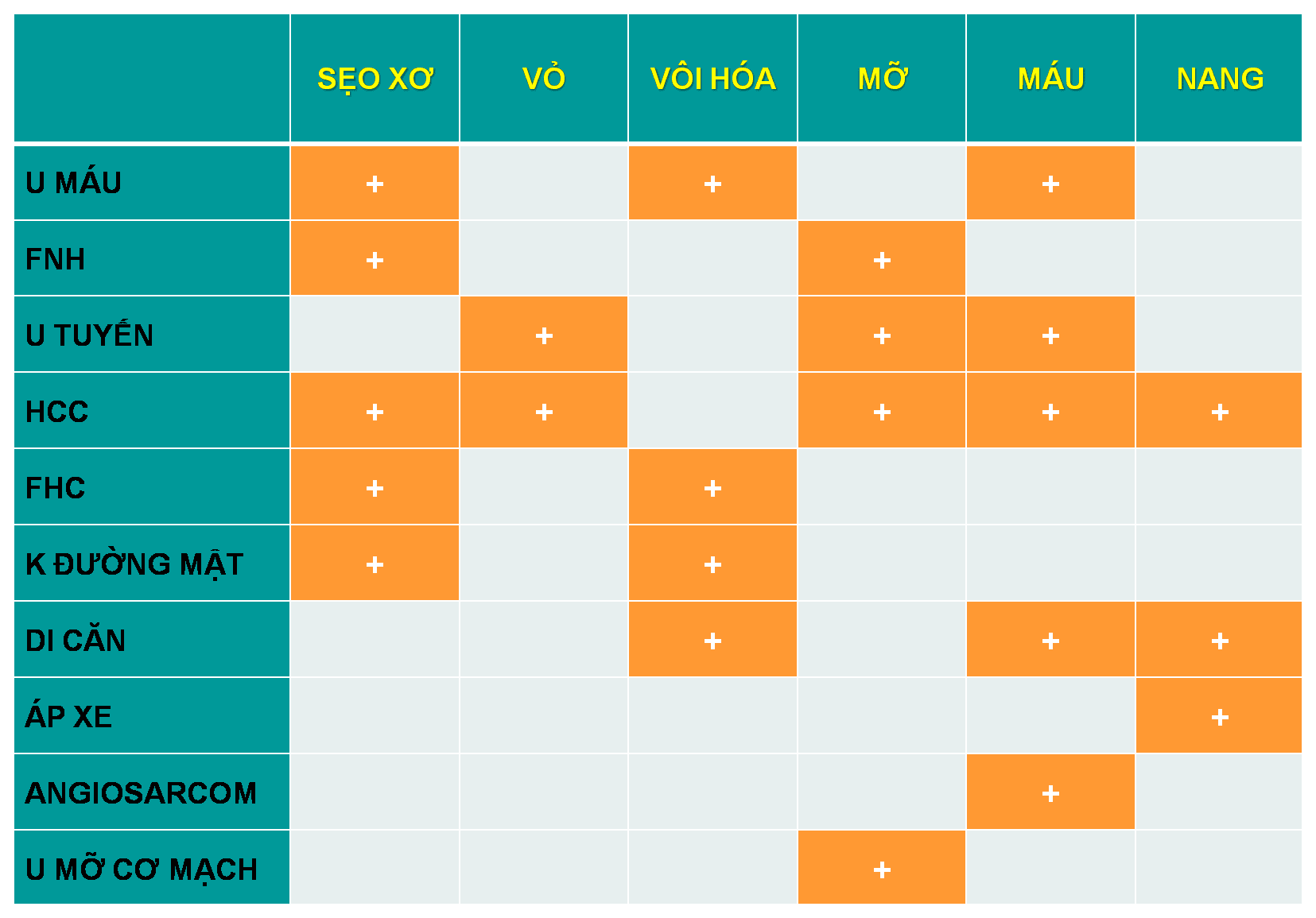
I. Đại cương
– U tuyến tế bào gan (Hepatic Adenoma) là tổn thương lành tính hiếm gặp, thường thường do hormone gây ra. Chúng bao gồm các bè gan không có ống dẫn mật hay khoảng cửa.
– Tỷ lệ tăng cao ở phụ nữ trong độ tuổi sinh đẻ dùng thuốc tránh thai đường uống Oestrogen trong thời gian dài, có thể thoái triển sau ngừng thuốc. Rất hiếm gặp ở nam giới trừ khi sử dụng steroid đồng hóa hoặc mắc bệnh dự trữ glycogen.
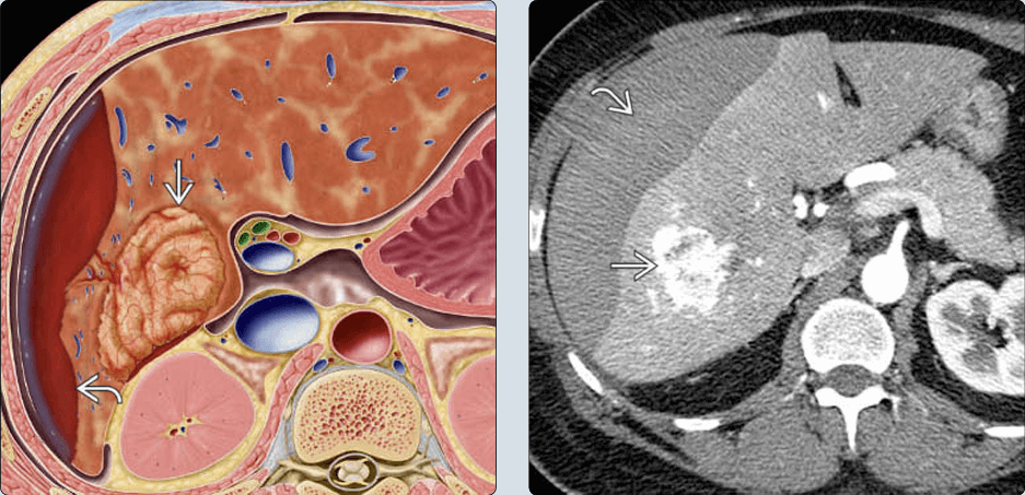
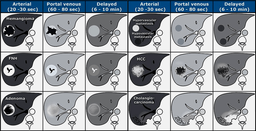
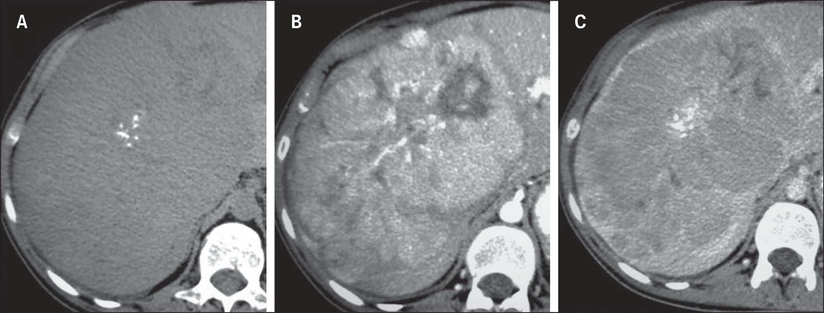
– Thường đơn độc, bờ giới hạn rõ, hình tròn, có vỏ giả bao bọc, kích thước lớn 8-15cm.
– Dạng đa ổ > 20% trường hợp. Khi có nhiều > 10 tổn thương gọi là u tuyến đa ổ (hepatic adenomatosis).
– Vị trí: thường ở dưới vỏ thùy gan phải (75%). Trong nhu mô hoặc có cuống (10%).
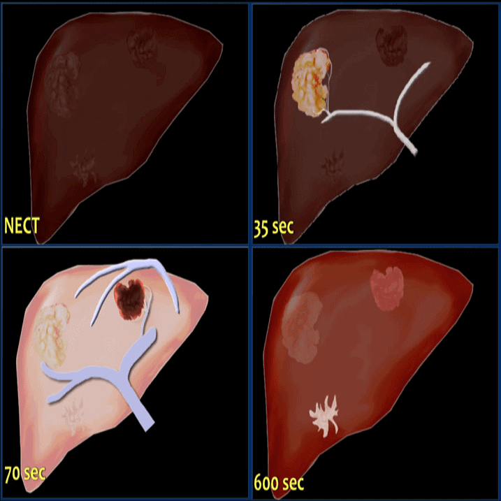
– U tuyến dễ xảy ra hoại tử trung tâm và chảy máu vì mạch máu nuôi bị giới hạn ở bề mặt khối u.
– Tiến triển: thường tiến triển rất chậm, có thể tăng kích thước nhanh trong trường hợp vẫn dùng thuốc tránh thai hoặc giai đoạn thai kỳ. Nguy cơ biến đổi ác tính ở 4,3% tổn thương > 5cm và vỡ tự phát kèm chảy máu đã được báo cáo ở 30% tổn thương > 5cm.
– Điều trị ngoại khoa khi có biến chứng: cắt bỏ, nút mạch gan chọn lọc.
– Phân loại: Dựa vào gen và giải phẫu bệnh:
+ Thể viêm (Inflammatory): phổ biến nhất, nguy cơ chảy máu cao. Liên quan đến béo phì và sử dụng rượu. Chủ yếu xảy ra ở phụ nữ.
+ Đột biến HNF 1 alpha: chủ yếu xảy ra ở phụ nữ, thường dùng thuốc tránh thai đường uống.
+ Đột biến beta catenin exon 3: nguy cơ cao ở nam giới dùng steroid đồng hóa. Nguy cơ cao nhất chuyển hóa thành HCC (47%).
+ Kết hợp viêm và đột biến beta catenin exon 3: chủ yếu mô học viêm. Phổ biến hơn ở nữ giới.
+ Đột biến beta catenin exon 7/8: nguy cơ chuyển hóa thành HCC không đáng kể. Nguy cơ chảy máu cao. Chỉ gặp ở bệnh nhân nữ. Liên quan đến bệnh dự trữ glycogen.
+ Kết hợp viêm và đột biến beta catenin exon 7/8: xhủ yếu mô học viêm. Liên quan đến béo phì và sử dụng rượu.
+ Sonic Hedgehog: nguy cơ chảy máu cao nhất. Liên quan đến việc sử dụng thuốc tránh thai lâu dài. Liên quan nhiều nhất với gan nhiễm mỡ.
+ Không phân loại: 5-10% các u tuyến không biểu hiện đặc điểm bệnh sinh hoặc phân tử đặc trưng.


Tài liệu tham khảo
* Focal Nodular Hyperplasia and Hepatic Adenoma: Differentiation with Low-Mechanical-Index Contrast-Enhanced Sonography – Diagnostic Accuracy of MRI in Differentiating Hepatocellular Adenoma From Focal Nodular Hyperplasia: Prospective Study of the Additional Value of Gadoxetate Disodium – Liver – Masses II – Common Tumors – Richard Baron
Hepatocellular Adenomas: Correlation of MR Imaging Findings with Pathologic Subtype Classification – Susanna M. van Aalten, MD, Maarten G. J. Thomeer, MD
* Hepatic Adenomas: Imaging and Pathologic Findings – Luigi Grazioli, MD, Michael P. Federle, MD, Giuseppe Brancatelli, MD
* Hepatic adenomatosis: MRI characteritics that can present like hepatocellular carcinoma – A. Sureshkumar, C. M. Valdes; Springfield, Massachusetts
* Hepatocellular adenoma: An unsolved diagnostic enigma – Matteo Renzulli, Alfredo Clemente, Francesco Tovoli
* MR Imaging of Hepatocellular Adenomas and Differential Diagnosis Dilemma – Luigi Grazioli, Lucio Olivetti, Giancarlo Mazza
* Current Approaches in the Management of Hepatic Adenomas – Diamantis I Tsilimigras, Amir A Rahnemai-Azar, Ioannis Ntanasis-Stathopoulos
* Hepatic adenomas: imaging and pathologic findings – L Grazioli, M P Federle, G Brancatelli
* Hepatic adenomatosis: spectrum of imaging findings – Matthanja Bieze1 Jacomina W. van den Esschert1 C. Yung Nio
* CT and MR Imaging Evaluation of Hepatic Adenoma – Giuseppe Brancatelli, Michael P Federle, Marie-Pierre Vullierme
* Hepatocellular Adenomas: Understanding the Pathomolecular Lexicon, MRI Features, Terminology, and Pitfalls to Inform a Standardized Approach – Maria Zulfiqar, Claude B Sirlin, Norihide Yoneda
* Hepatocellular adenoma: imaging review of the various molecular subtypes – H. Dharmana, S. Saravana-Bawan, S. Girgis, G. Low
* Long-term follow-up of hepatic adenoma and adenomatosis: analysis of size change on imaging with histopathological correlation – N Shao, A Pandey, M A Ghasabeh
* Hepatocellular adenomas: magnetic resonance imaging features as a function of molecular pathological classification – Hervé Laumonier, Paulette Bioulac-Sage, Christophe Laurent
* Inflammatory hepatic adenomas: Characterization with hepatobiliary MRI contrast agents – James F Glockner, Christine U Lee, Taofic Mounajjed
* Genetics and imaging of hepatocellular adenomas: 2011 update – Venkata S Katabathina, Christine O Menias, Alampady K P Shanbhogue
* New MRI features improve subtype classification of hepatocellular adenoma – Sylvain Bise, Nora Frulio, Arnaud Hocquelet, Nicolas Alberti
* Radiology Illustrated Hepatobiliary and Pancreatic Radiology – Byung Ihn Choi
# Cập nhật nội dung bài viết & Case lâm sàng 23/3/2024
# Cập nhật nội dung bài viết & Case lâm sàng 8/3/2024
# Cập nhật nội dung bài viết & Case lâm sàng 6/2/2024
# Cập nhật nội dung bài viết & Case lâm sàng 15/10/2023