I. Kỹ thuật
* Thì trước tiêm
– Thì trước tiêm (non enhanced CT – NECT)
– Mức độ phát hiện của 1 tổn thương gan phụ thuộc vào sự khác biệt tỷ trọng giữa tổn thương và nhu mô gan bình thường xung quanh.
– Trên phim chụp cắt lớp vi tính không tiêm (NECT), khối u gan thường khó quan sát thấy vì độ tương phản giữa mô u và nhu mô gan xung quanh quá thấp.
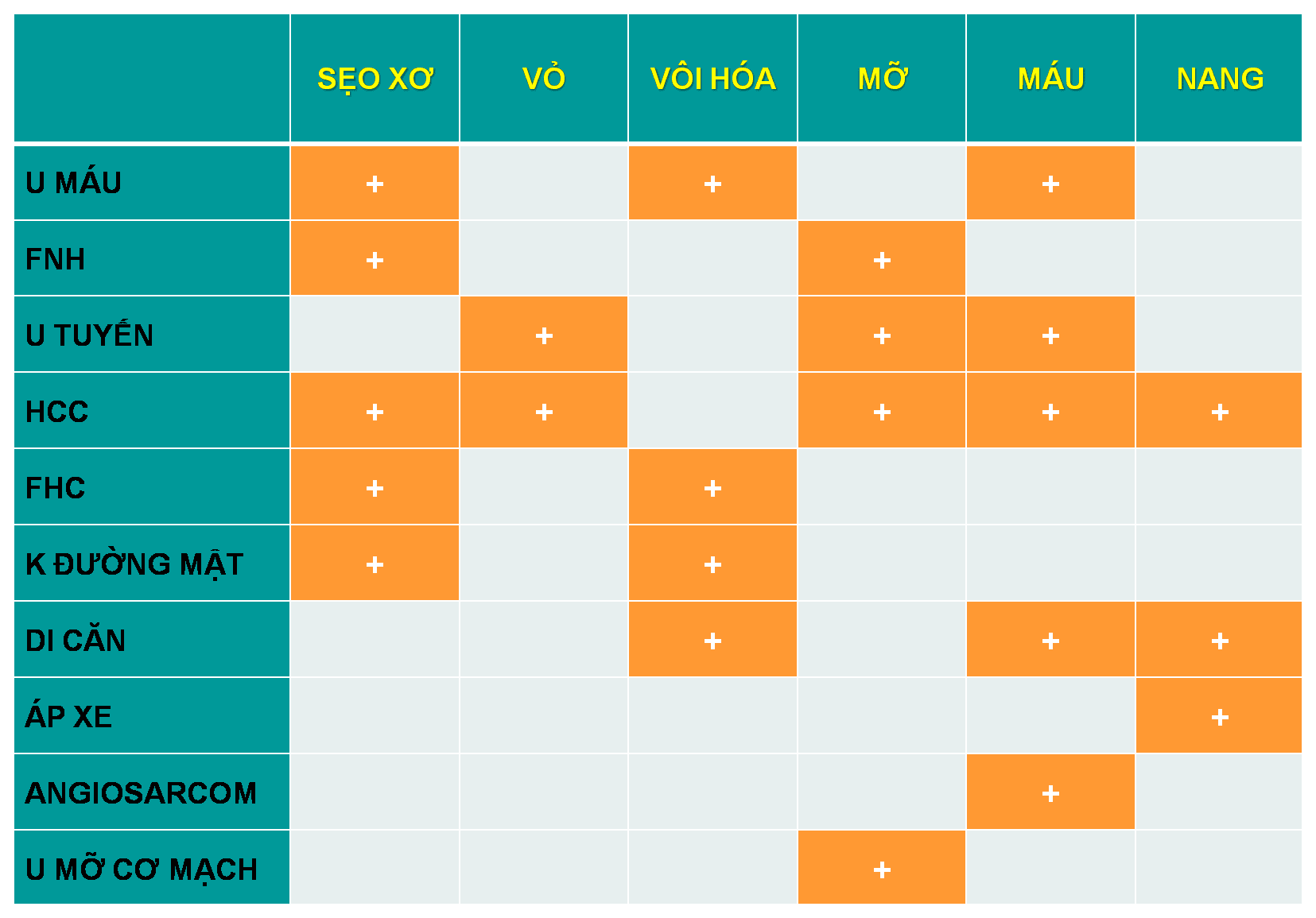
– Một số ít khối u chứa vôi hóa, thành phần nang, mỡ hoặc xuất huyết sẽ được phát hiện trên phim chụp không tiêm thuốc cản quang => Vì vậy, tiêm thuốc cản quang là cần thiết để phát hiện 1 tổn thương gan.
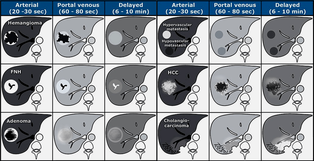
– Khi tiêm thuốc cản quang điều quan trọng cần hiểu là gan có một nguồn cấp máu kép: nhu mô bình thường được cung cấp 80% bởi tĩnh mạch cửa và chỉ 20% bởi động mạch gan => vì vậy nhu mô gan sẽ ngấm thuốc trong thì tĩnh mạch cửa.
– Tuy nhiên, tất cả các khối u gan đều nhận được 100% nguồn cung cấp máu từ động mạch gan, vì vậy chúng sẽ ngấm thuốc ở thì động mạch.


Tài liệu tham khảo
* Characterisation of liver masses – Richard Baron
* A practical diagnostic approach to hepatic masses – Monika Vyas
* Diagnostic Approach to Hepatic Mass Lesions and Role of Immunohistochemistry – Esmeralda Celia Marginean
* Focal Hepatic Lesions: Diagnostic Value of Enhancement Pattern Approach with Contrast-enhanced 3D Gradient-Echo MR Imaging – Khaled M. Elsayes
* Imaging Approach for Evaluation of Focal Liver Lesions – Daniele Marin
* Approach of the patient with a liver mass – Carlos Rodríguez de Lope
* Cystic Hepatic Lesions: A Review and an Algorithmic Approach – Amir A. Borhani
* Approach to the Solitary Liver Lesion: Imaging and When to Biopsy – Emily H.T.PangMD, FRCPCAlison C.HarrisMD, FRCPCSilvia D.ChangMD, FRCPC
* Liver imaging reporting and data system and CT/MRI diagnosis of hepatocellular carcinoma – Devaraju Kanmaniraja , Victoria Chernyak
* Bleeding Liver Masses: Imaging Features With Pathologic Correlation and Impact on Management – Aaron J. Thomas
* Diagnostic approach to hepatic hemangiomas – R C Nelson, J L Chezmar
* LI-RADS Liver Imaging Reporting And Data System – Frederieke Elsinger
* Imaging pitfalls in MDCT of hepatocellular carcinoma: an imaging collection of lessons learned – K. M. Wong, A. C. C. Poh, A. G. S. Tan; Singapore/SG
* Pitfalls in CT and MRI differential diagnosis of focal liver masses with central scar – S. Ghiea, M. Boros, C. Dobromir, I. Lupescu, S. A. Georgescu; Bucharest/RO
* Hepatic lesions with capsular retraction: a diagnostic challenge – V. M. Vilela, A. M. Mota Mustafá, M. Vinicius Bastos Sasaki, W. Paula, M. V. A. Soares; Brasilia/BR
* CT and MR findings of the various causes of hepatic capsular retraction: A pictorial review – D. M. Yang, H. S. Kim, J. H. Kang, C. Park; Incheon/KP
* The many faces of Hepatocellular carcinoma: An analysis of 139 cases of hepatic mass lesions – A. Malhi
* Fat-containing Lesions of the Liver: Radiologic-Pathologic Correlation – Srinivasa R. Prasad, Hanlin Wang, Humberto Rosas, Christine O. Menias, Vamsi R. Narra, William D. Middleton, Jay P. Heiken
* Fat-Containing Liver Lesions on Imaging: Detection and Differential Diagnosis – Andreu F. Costa
* Liver Masses With Central or Eccentric Scar – Tonsok Kim, MD, PhD, Masatoshi Hori, MD, PhD, and Hiromitsu Onishi, MD, PhD
* Liver Calcifications and Calcified Liver Masses: Pattern Recognition Approach on CT – Madhavi Patnana
* Imaging of calcified hepatic lesions: spectrum of diseases – Giuseppe Mamone
* Liver capsule retraction: a pictorial review – C. Bilreiro, C. Carneiro, J. Saraiva, J. Brito; Portimão/PT
# Cập nhật nội dung bài viết & Case lâm sàng 12/6/2026